Research Article - Clinical Schizophrenia & Related Psychoses ( 2023) Volume 17, Issue 4
Novel Hypotheses on the Role of Microglia and PANoptosis in Neurocysticercosis
Lourdes de Fatima Ibanez Valdes and Humberto Foyaca Sibat*Humberto Foyaca Sibat, Department of Neurology, Nelson Mandela Academic Central Hospital (NMACH), Walter Sisulu University, Mthatha, South Africa, Email: humbertofoyacasibat@gmail.com
Received: 08-Sep-2023, Manuscript No. CSRP-23-11324; Editor assigned: 11-Sep-2023, Pre QC No. CSRP-23-113249 (PQ); Reviewed: 26-Sep-2023, QC No. CSRP-23-11324; Revised: 03-Oct-2023, Manuscript No. CSRP-23-113249 (R); Published: 10-Oct-2023, DOI: 10.3371/CSRP.DLHF.101023
Abstract
Background: Cysticercosis (Ct) is a preventable and eradicable zoonotic parasitic disease secondary to an infection caused by the larva form of pig tapeworm Taenia solium (Ts), mainly seen in people living in developing countries. However, the number of carriers in developed countries has increased gradually due to globalization and uncontrolled migration. In this study, we look for the role Microglia (Mg) played in the pathogenesis of Neurocysticercosis (NCC). After viewing this issue, we formulate some hypotheses regarding the role of Mg, trained immune response and PANoptosis in NCC.
Methods: We searched the medical literature comprehensively, looking for published Medical Subject Heading (MeSH) terms like "Neurocysticercosis", "pathogenesis of neurocysticercosis", "comorbidity in NCC", OR "Apoptosis", "Pyroptosis," OR "Necroptosis;" OR "PANoptosis;" OR "PANoptosome;" OR "Reprogramming Somatic Cells."
Results: All selected manuscripts were peer-reviewed, and we did not find publications related to Mg/Ap/Pp/Np/PANp/PANop/RSC/NCC.
Conclusion: We have hypothesized the role of Mg on the pathogenesis of cysticercus perilesional oedema and the role of Mg during NCC's colloid/nodular stage.
Keywords
Cysticercosis • Neurocysticercosis • Microglia activation • Apoptosis • Pyroptosis • Necroptosis • PANoptosis • PANoptosome
Abbreviations
Aβ: Amyloid-β; AD: Alzheimer’s Disease; AIDS: Acquired Immune Deficiency Syndrome; AKT: protein Kinase B; ALS: Amyotrophic Lateral Sclerosis; CaMKII: Calcium-calmodulin Kinase II; c-FLIP: cellular FADD-Like Interleukin-1β converting enzyme inhibitory Protein; cIAP1: cellular Inhibitor of Apoptosis Protein 1; CNS: Central Nervous System; ADRs: Activated Death Receptors; ERK1/2: Extracellular signal-Regulated Kinases 1/2; FADD: Fas-Associated Death Domain; FPI: Fluid Percussion Injury; GSK3: Glycogen Synthase Kinase 3; IKK: IκB Kinase; IRI: Ischemia-Reperfusion Injury; IRI: Ischemia-Reperfusion Injury; LPS: Lipopolysaccharide; MLKL: Mixed Lineage Kinase Domain-Like Protein; Nec-1: Necrostatin-1; NEMO: Nuclear factor-kappa B Essential Modulator; NLRP3: NLR family Pyrin domain containing 3; OPTN: Optineurin; PCD: Programmed Cell Death; PD: Parkinson’s Disease; PI3K: Phosphorylates and activates phosphoinositide 3-Kinase; PD-1: Programmed cell Death-1; PKC: Protein Kinase C; RIC: RIPK1-Inhibitory Compound; RIPK1: Receptor-Interacting Protein Kinase 1; RIPK3: Receptor-Interacting Protein Kinase 3; sALS: sporadic Amyotrophic Lateral Sclerosis; SN: Substantia Nigra; SOD1: Superoxide Dismutase 1;TAK1: Transforming growth factor β-Activated Kinase-1; TBI: Traumatic Brain Injury; TICAM-1: TIR domain-Containing Adaptor Molecule 1; TLR: Toll-Like Receptor; TNF: Tumor Necrosis Factor; TNFR1: TNF Receptor 1; TRADD: TNFR-Associated Death Domain; TRIF: Toll/IL-1 Receptor domain-containing adaptor inducing IFN-β; WNV: West Nile Virus; WT: Wild-Type.
Introduction
Cysticercosis (Ct) is a preventable neglected zoonosis but eradicable parasitic disease secondary to a cestode infection by the larva form of the pig tapeworm Taenia solium (Ts), most often seen in people living in developing countries. Ct can fest any internal organ in humans and pigs, including the hair, nails, bone tissue, epidermis, cartilage, and the adrenal gland. When the cysticercus is in the cerebral parenchymal, intraventricular system, Subarachnoid Space (SAS), cerebellum, brainstem, optic nerve, or spinal cord, then it is best known as Neurocysticercosis (NCC), and the most common clinical manifestations include headaches and epileptic seizures/epilepsy among other less frequent symptoms and signs [1-5]. Epileptic Seizure (ES) disorder and Epilepsy (Ep) are the most common symptom of Intraparenchymal NCC (INCC). We performed more than ten epidemiological investigations in rural areas around Mthatha (South Africa), confirming that NCC is the leading cause of secondary epilepsy. All ES, and Ep, respond very well to first line Antiseizure Medication (ASM) and Antiepileptic Drugs (AED) [6-10]. Likewise, lack of available AED due to COVID-19 restrictions or other reasons, including financial constrictions and poor compliance, leads to Status Epilepticus (SE) complications. Despite this, patients presenting with refractory epilepsy secondary to NCC without other causes have not been seen in our region over the past 26 years [11-15]. The most used ASM is benzodiazepine, and the most common AEDs include valproic acid and carbamazepine. Levetiracetam is used only in tertiary hospitals because it is not available in our rural areas [16-19].
Humans are the final host for the adult tapeworm (taeniasis). In contrast, humans and pigs can be intermediate hosts carrying the cysticercus (larval form), a cyst, fluid-filled membrane vesicles with an eccentric scolex inside. When the cysts are ingested by undercooked contaminated pork meat, it goes to the gut, where scolex evaginates. It becomes attached to the intestinal mucosa wall by two crowns of hooks, avoiding expulsion out of the intestine by peristaltic movement. At the gut, one or a maximum of two parasites matures into a 2-4 meters length tapeworm, constituted by a scolex, neck and 1,200 proglottids. Gravid proglottids contain from 600 to 2000 fertile eggs, which pass into the soil after defecation on alternating days if the person is not constipated or has diarrhea [16-19]. In impoverished countries or economically poor regions inside of advantageous countries (like our area) where access to clean and safe water is not possible and predominates poor sanitation, poor food/personal hygiene, poor educational health, high level of poverty and free-roaming pigs with access to human faeces contaminated by Ts eggs, the incidence/prevalence of NCC is notably high. When the proglottids or eggs are ingested by contaminated water, food or via the fecal-oral route, the embryos are released from the egg into the gut and pass through the gut mucosa to the blood flow, which carries them to the target tissues, where they are transformed into cysticerci. Like human beings, pigs can ingest eggs and develop porcine cysticercosis [20-26]. Person-to-person transmission is relatively standard and explains how non-eaten pork peoples are infected and why the disease is present in developed countries without free-range pigs and even in places where the four stages of cysticercus in the brain parenchymal have been identified [12].
Recently, we reviewed several novel aspects of NCC associated with COVID-19 and Human Immunodeficiency Virus (HIV), the autoimmunity, meningeal lymphatic, glymphatic drainage, and the role of activated Oligodendrocyte/Oligodendrocyte Precursor Cell (OLG/OPC/NG2) in the pathogenesis of NCC clinical manifestations/complications/outcome. As we commented previously, activation of microglia and astrocytes is at the centre of NCC neuroinflammatory pathways either directly or indirectly due to their secretion of pro-inflammatory cytokines, upregulation of Blood-Brain-Barrier (BBB) disrupting proteinases and formation of an inhibitory glial scar [27-29].
In 2006, we commented on the clinical features and other aspects related to Spinal Cord NCC (SCNCC) [28], and recently we commented on the role played by pericytes on the pathogenesis of local neuroinflammation due to NCC, the healing process and outcome of the SCNCC [29]. Most glial cells support the primary function of the Central Nervous System (CNS), including maintaining homeostasis of Neurons (NC)/Glial Cells (GC). In addition, as is well-known that most Astrocytes (Ast), Microglia (Mg), OLD, Schwann cells, and ependymal cells are capable of mitotic division. We have recently commented on the OLG's structure and role in NCC [19]. The Mgs are involved in developing brain tissue structure, phagocyting/removing waste metabolite, foreign/damaged organisms, material, or cells [30].
Most researchers considered the existence of two types of Mgs known as M1, which release toxic substances and inflammatory factors to inhibit pathogens, and M2, which exerts neuroprotective functions by promoting restoration of impaired immune responses and subsequent regeneration and nerve repair. In 1919, the Spanish neurologist Pio del Rio Hortega, also known as a "Father of Microglia", proposed the existence of Mg subtypes. However, it was ignored until 2016, when his ideas were retaken. Nonetheless, it has been universally accepted that the Spanish scientist first described Mgs as a new type of GC produced in the yolk sac by Mp (Macrophage) migration from the periphery into the CNS around 10.5 days of embryonic development. He also found that the cell body of some Mg was associated and interacted with the initial segment of the axons and called it "satellite" Mg, where the action potential starts [31].
The main aim of this comprehensive review is to answer the following research questions. 1. What is the role of Mg in NCC according to published information? 2. What’s the role of PANptosis in NCC? 3. What is the role of somatic cell reprogramming in NCC? Moreover, what is the role of trained immunology in NCC?
Materials and Methods
A systematic search of EMBASE (RRID: SCR 001650), MEDLINE RRID: SCR 002185), Cochrane Library (RRID: SCR 013000), Scopus (RRID: SCR 022559), PsycINFO (RRID: SCR 014799), and EBSCO CINAHL (RRID: SCR 022707) was conducted to identify articles published between January 31st, 2003, to January 31st, 2023, followed by hand-searching of relevant journals.
Literature search strategy for this study
These databases support the comprehensive search of various topics in health and healthcare: We screened all papers regarding the issues mentioned earlier from the primary and secondary health care and research centre under the search terms "NCC," "Mg activation", and "Ap, Pp, Np, PANp, PCD".
We selected for study those that were relevant to these issues.
For practice guidelines, we reviewed the references of each included manuscript.
The aim is to select the original research studies related to the search mentioned above strategy.
After that, the search was restricted to full-text written in Spanish, Portuguese, and English language. All studies were retrieved using MeSH, only including aspects within the current work scope.
Inclusion and exclusion criteria
We also selected randomized controlled trials or quasi-experimental studies published in peer reviewed journals. However, the studies were excluded if they evaluated interventions for other types of parasitic infections, other infections, or vascular problems because their etiologies differ from T solium infection. In addition, the study was also limited to studies involving adult patients and published in English.
Study selection
We performed the literature search and scanned all articles by title and abstract. LdeFIV and HFS independently screened articles for eligibility. It was followed by a discussion to establish consensus on which studies were included, mainly when there was ambiguity.
Quality appraisal
Four areas of study quality were assessed: selection bias, study design, health status, blinding process, reasons for dropouts or withdrawals, and data collection methods. In addition, LdeFIV independently carried out a methodological quality assessment, which HFS verified.
Data extraction
A data extraction mechanism was developed to extract research data about the setting, study design, demographic profile of patients, methods, measurement tools and timing of assessments, and outcomes when met the inclusion criteria. In addition, crucial information was extracted from either the primary article or an earlier published manuscript on the intervention for secondary data analysis studies. HFS conducted the data extraction; the consensus was achieved through discussion among other colleagues.
Methods of analysis
Extracted data were initially synthesized using textual descriptions to determine the characteristics of the selected studies, and then they were grouped, clustered, and presented in tabular form.
Study and cohort selection
We selected prospective and retrospective case reports, cross-sectional studies, cohort studies, case-control studies, case series, reviews, controlled clinical trials, and meta-analyses releasing data on inclusion criteria.
Data collection process
The selected information was extracted from each manuscript with Microsoft Excel (RRID: SCR 016137) in a structured coding scheme. The data collected included HIV-NP, HHV7/HIV disorders, clinical features, population size, age distribution, and the investigations used to confirm the final diagnosis when applicable. In cases where there was uncertainty regarding the interpretation of the selected data or how it could be used, we analyzed the situation until we arrived at a mutual agreement.
Quality assessment of selected publications
Initially, all studies were screened for bias using the Jadad scoring system as usual and included only those with Jadad scores ≥ 4 for further assessment.
Effect measures and certainty assessment
The risk ratio/mean difference/confidence procedures used in the synthesis were applied to publications that met the inclusion criteria only.
Results
All selected manuscripts were peer-reviewed publications, and none met all inclusion criteria on Mg/Mp/Ap/Pp/Np/PANp/PANop/trained immunology/and RSC.
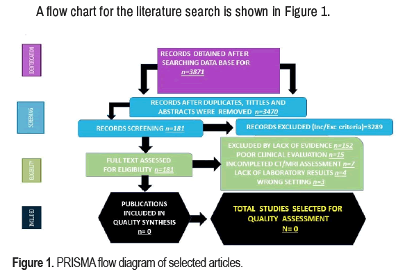
A flow chart for the literature search is shown in Figure 1.
Study characteristics
Ethics committees approved all publications included in this study, and patient consent was obtained; without this requirement, the pertained data were removed from this study. The number of people with NCC ranged from 45,342 to 49,671 (median 47,506.5 Interquartile Range (IQR) 12,602–36,610). In South Africa, 49.44% of the population was HIV-positive, and 59.2% were female. Most investigations were conducted in the United States of America/Canada (42.1%), followed by Asia (39.5%), Africa (12.7%) and European countries (5.7%). Most studies (77.3%) focused on people older than 18. The total number of publications identified was 3,871; after duplicate removal, n=3,470; after full text excluded, n=181; for quality synthesis, n=0; for quality assessment, n=0.
Discussion
Most studies combined cases-series, case reports, immunological analyses, and medical literature reviews. Most affected populations were probably not included in those reports because of the need for proven diagnostic confirmation. In addition, due to a scarce number of studies in children, most clinical features, demography, and immune response still need to be included.
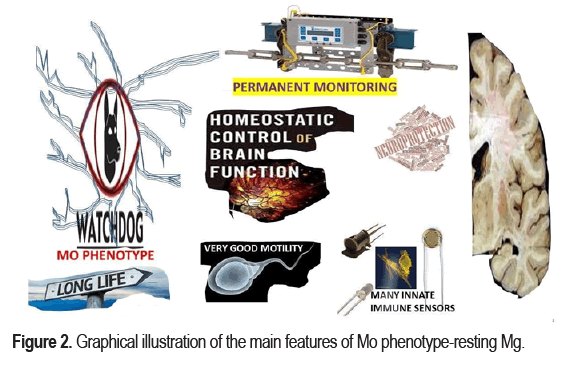
We identified four stages of cysticercus in the brain, known as I-Vesicular: viable parasite with intact membrane, no-host immunological reaction, and no local Neuroinflammation (NI). II-Colloidal: the dying process of the parasite commonly before five years of entry. The cyst fluid becomes turbid. Compared with the CSF density, it is the darkest. The damaged membrane leaky oedema surrounds the cyst. In this stage, the neurological manifestations are more evident due to the direct/indirect effects of the released parasite's antigen. III Granular/ nodular: decreases surrounding perilesional oedema, and the cyst begins to retract, but the enhancement persists. IV) Calcified: Sometimes residual perilesional oedema, all structural characteristics of the cyst disappear, and the remnant material is calcified [29,31]. As we documented before, activation of Microglia (Mg)/Astrocytes (Ast) and Pericytes (Pc) are at the centre of NCC neuroinflammatory pathways either directly or indirectly due to their secretion of pro-inflammatory cytokines, upregulation of BBB disrupting proteinases and formation of an inhibitory glial scar [19,29]. We will view aspects related to Mg/IS/ES/Ep and oxidative stress related to NCC soon; now, we will comment on the relationship between Mg and NCC only due to the constriction of many pages for this publication. The Mg first proliferates, extending the length of their nuclei, forming aggregates around the necrotic tissue, or dying cell bodies and releasing Reactive Oxygen Species (ROS) like P2X7, CB2 and COX-2, which are involved in the etiological mechanism of Multiple Sclerosis (MS) and Amyotrophic Lateral Sclerosis (ALS) as we commented before [32]. As aforementioned, Mgs are the only resident innate immune cells derived from the nerve tissue's mesodermal layer. Apart from the homeostatic functions, preventing overshooting immune responses during development, health, and disease because they are perfectly anatomically ubicated to act as accurate tissue watchdogs, favored by their long life, remarkable good motility, and abundant immune sensors. They also are involved in the development, maturation, repair, and endogenous immune response by neuroprotective/ neurotoxic actions of the CNS.
Classically, the Mgs were considered in the permanent resting state (MO phenotype) under neurophysiological conditions. Their main functions are surveillance of the CNS immunologically by permanently monitoring all pathological events in the CNS, maintaining a relatively quiescent neuron-specific monitoring phenotype, and playing an "immune surveillance and defence" role in the microenvironment of nerve cells as shown in Figure 2.
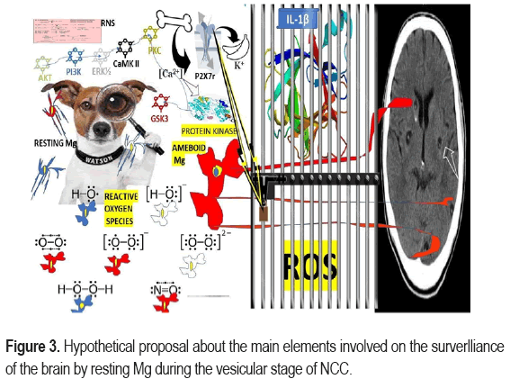
Furthermore, they can extend out of the cell body, constantly monitoring for any potential damage in the brain in response to an informative signal from the surrounding cells. Therefore, Mg performs a complete assessment of the CNS for any injury/damage/infection every few hours and modulates the necessary investigations to determine the immune status of the brain continuously. Nonetheless, it has been proposed that the MO implement functional and structural changes under pathological conditions and polarizes into the typical M1 (activated Mg) or M2 alternatively. Therefore, they rapidly activated and polarized, accompanied by a series of events such as phagocytosis, proliferation, chemotaxis, migration, and pro-inflammatory cytokine production such as Interferon-γ (IFN-γ), Interleukin-1β (IL-1β), Tumour Necrosis Factor-α (TNF-α), proteolytic enzymes, including Heme Oxygenase 1 (HO-1), Matrix Metalloproteinases (MMPs), a large number of reactive oxygen species, inducible Nitric Oxide Synthase (iNOS), and Interleukin 12 (IL-12) an adaptive immune response, remove foreign pathogens, and a sustained inflammatory response aggravating brain damage as shown in Figure 3.
Reactive Nitrogen Species (RNS), are a family of antimicrobial molecules which main source is Nitric Oxide (NO) and superoxide (O2•−). Both are produced through the enzymatic activity of NADPH oxidase and Nitric Oxide Synthase 2 (NOS2) respectively, Reactive Oxygen Species (ROS) surge from biochemical reactions that occur during respiration and photosynthesis in mitochondria, peroxisomes and chloroplasts. It’s well known that mechanism of ATP production in the mitochondria (oxidative phosphorylation), include transport of protons across the inner mitochondrial membrane to oxidation-reduction reactions. Included P2X7, CB2, COX2.
During the respiration process the mitochondria convert energy for the cell into a usable form, Adenosine Triphosphate (ATP). The process of ATP production in the mitochondria, called oxidative phosphorylation, involves the transport of protons (hydrogen ions) across the inner mitochondrial membrane by means of the electron transport chain. P2X7=P2X Purinoceptor 7 is a protein belongs to the family of purinoceptors for ATP. The receptor is commonly found in the CNS/PNS, Mg/Mp, uterine endometrium, and in the retina. Ideally, concentrations of ATP will activate a homotrimer protein cation channel P2X7 receptor. In cases of NCC, we speculated that after a prudential expression time, it complexes with membrane proteins create a wide pore at the cellular membrane that leads to increased released of ATP into the extracellular milieu and PCD. Other authors have established that P2X7 receptors are widely expressed in the CNS Mg mainly at the level of hippocampus, amygdala, frontal cortex, and striatum, regions involved in neurodegenerative diseases and psychiatric disorders. We have hypothesized that P2X7 receptor is highly expressed in NCC Mg where mediated CP/PCD, fast and reversible membrane blebbing, multinucleated cell production of exosomes, phosphatidylserine exposure, release of microparticles formation of nitrogen species and ROS. P2X7r expression also triggers intracellular signalling pathway and binding ATP modify its state and open membrane pore for the entrance of Ca2+ plus other cations. In case of NCC we speculate that the elevated concentration of intracellular Ca2+ ((Ca2+) i) activate some kinases (a protein enzyme that increase speeds of chemical reactions) such as, CaMKII, PKC, PI3K, AKT, GSK3, and ERK1/2 leading to inhibition of Ap or elevate genetic transcription of cell survival. In Figure 3, we also included (as part of this hypothesis) the consequences of activation of P2X7r releasing IL-1β/ROS from “jail” and increasing inhibition of GSK3 if the formation of the NLRP3 inflammasome and NF-κB are completed. Finally, we have hypothesized that administrating P2X7 receptor antagonists in patients with massive NCC instead of PZQ/ALB/St may provide some improvement of NI without risk of development of ES/SE and death.
M1 Mg is a pro-inflammatory cell that serves as the first line of defence of the neuroimmune system arriving at the scene in a few hours after an appropriate stimulation and generating complex responses according to the intensity of the brain injury sending. In mild injury, the M1 sends a "localize me" signal followed by M2 state activation characterized by small cell bodylike changes, distal branches, secretion of anti-inflammatory elements, cell tissue regeneration, nerve repair, phagocytic activity, and neuroprotective roles. In cases of intensive lesion/damage, the Mg sends a "swallow me" signal followed by an activated toxic M1/pro-inflammatory state, typically characterized by amoeboid modifications characterized by thick protrusions and large and round cell bodies able to remove debris of dying cells, infected cells and exert cytotoxic effects. Nevertheless, exaggerated M1 activation leads to neuronal dysfunction and cell loss/damage/degeneration. These M1/M2 Mg conduct different activities responding to the inflammatory stages and environmental stimulations. Nucleotide oligomerization domain receptors, toll-like receptors, and scavenger receptors recognize the harmful stimulus. An endorsed level of M1 Mg has been reported in response to Lipopolysaccharide (LPS), other bacterial components or signals of CNS infections if IFN-γ is elevated.
A brief comment about M1 phenotype in NCC
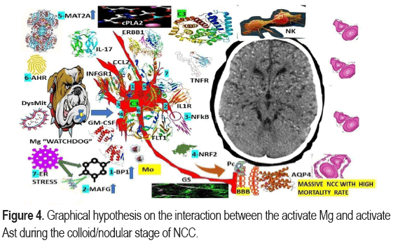
In cases of the sterile inflammatory response caused by CNS ischemic/reperfusion/trauma, M1 from depolarized Mg reduces Nicotinamide Adenine Dinucleotide Phosphate (NADPH), secretes chemokines, Interleukin-6 (IL-6), Interleukin-1α (IL-1α), and IL-12 plus surface antigen CD40 plus other elements with the capacity of inhibit phagocytosis if the production of NLRP3 inflammasome, ROS or iNOS fail. Supported by the information mentioned earlier, we hypothesize that in patients presenting with NCC at the colloid stage in response to released antigens from the dying process of cysticerci, then the activated M1 Mg secrete the cited pro-inflammatory elements, participate in the process of clearance of waste metabolite, and reduce the intensities of the inflammatory process at the pericystic area but according to the quality/quantity of the parasitic lesion then M1 phenotype can promote NI and inhibit phagocytosis causing neuronal irritation/injury/damage triggering ES/Ep at the cortical level and causing headache by subsequent irritation of the nearby trigeminal receptor in the dura matter. Later, t is an overreaction of M1 Mg leads to increased NI and a delayed nerverepairing process. The path genic process is more complex if associated ischemia/reperfusion happens as shown in Figure 4.
We have hypothesized that because the closed proximity between Mg and Ast, the second supportive cell to be activated in the brain, even before the immune elements located at the border of the CNS at the meninges/choroid plexus. The mechanism of swift As’ responding to the Mg signals in humans have not been proved up to date, but we are building up some hypotheses based on animal investigation’s reports. Other authors have documented that Ast’ surface receptor such as: FLT1, ERBB1, IL1R, TNFRs, GM-CSFRs, IL-17R, and IFNGRs are the main mediators in this process. The participation of XBP1 which is activated by endoplasmic reticular stress in also illustrated in Figure 4 as another component of this hypothesis, plus elevation of MAFG, MAT2A and C3 intracellularly. We also speculate that activate Ast act as a direct respond of activated/M1/ Mg increase their secretion of proinflammatory molecules as we commented under other circumstances but now including CCL2/GM-CSF leading to recruitment of encephalitogenic T-lymphocytes plus other proinflammatory monocytes causing NI at the pericystic colloid/nodular NCC [2]. Other elements involved in this hypothesis are cPLA2, and NK as has been illustrated in Figure 4. FLT1 is a member of VEGF receptor gene family, encodes a receptor tyrosine kinase which is activated by VEGF-A, VEGF-B, and placental growth factor ERRB1. The epidermal growth factor receptor (also known as EGFR; ErbB-1; HER1) is a transmembrane protein receptor for members of the epidermal growth factor family (EGF family) of extracellular protein ligands, Interleukin 1 Receptor (IL1R), type I also named as CD121a, is an interleukin receptor. Interleukin 17 (IL-17) family is a family of pro-inflammatory cystine knot cytokines, produced by a group of T helper cell named as T helper 17 cell, Interferon Gamma Receptor 1 (IFNGR1) also called as CD119, is a protein encoded by the IFNGR1 gene, cytosolic Phospholipase A2 (cPLA2). The chemokine (C-C motif) Ligand 2 (CCL2) is also named as Monocyte Chemoattractant Protein 1 (MCP1) and small inducible cytokine A2 and it is a small cytokine that tightly modulates cellular mechanics and thereby recruits memory T cells, monocytes, and dendritic cells to the sites of NI produced by the colloid/nodular stage of NCC according to our hypotheses, (1) X-box Binding Protein 1 (XBP1), also called as XBP1, is a transcription factor protein which regulate genes expression to support the immune system and the cellular stress response, (2) Transcription factor MafG is a bZip Maf transcription factor protein is one of the small Maf proteins, which are basic region and leucine Zipper (bZIP)-type transcription factors, (3) Nuclear Factor Kappa-light-chain-enhancer protein of activated B cells (NF-kB) control cytokine production, and transcription of DNA, and its involved in cell survival, cellular responses to stimuli such as stress, free radicals, heavy metals, cytokines, ultraviolet irradiation, oxidized LDL, and bacterial/viral antigens and we assumed parasitic antigens too. NF-κB also participate in regulating the immune response to infection, Complement component 3 (C3), is a protein of the immune system in the blood and plays a remarkable role in the complement system contributing to innate immunity. (4) The Nuclear factor erythroid 2-Related Factor 2 (NRF2) is a RNA element present in the 5′ UTR of the mRNA encoding the transcription factor Nrf2 and an emerging regulator of cellular resistance to oxidants, Methionine Adenosyl Transferase (MAT2A) also known as S-adenosylmethionine synthetase is an enzyme that creates S-adenosylmethionine and is also involved in cell proliferation, gene transcription, and production of secondary metabolites. The Aryl Hydrocarbon Receptor (AHR) (also known as ahR, AhR, ahr, AH receptor, or dioxin receptor) is a protein transcription factor that regulates gene expression. The Blood-Brain Barrier (BBB) is a remarkable selective semipermeable border of EC which ontrol that solutes in the brain circulation do not cross into the extracellular fluid of the CNS to protect neurons while allow the entry of the necessary nutrients, Corpora Amylacea (CA) is a small hyaline masses found in the lungs, the nervous system, and the prostate gland, among other organs of the body which increase in advancing age, and its involved in the clearance the metabolite waste in the CNS. Aquaporin-4, is a water channel protein belongs to the aquaporin family of integral membrane proteins that conduct water through the cell membrane and participate in the clearance system of the brain. The Glymphatic System (GS) oversees waste clearance in the brain of vertebrates modulating the flow of the CSF into the paravascular space around blood vessels mixing the interstitial fluid and parenchymal solutes and exiting down CNS venous paravascular spaces with the capacity to protect the neural network. Unfortunately, we have not evidence how the clearance system works at the DRG (apart from the fenestrate CV) and at the enteric nervous system (apart from gut/brain axis/CLN). Pericytes (Pc) are only cell located between endothelial cells, astrocytes, and neurons within the neurovascular unit extending their processes along capillaries, pre-capillary arterioles and post-capillary venules. Pc contact with more than 90% of the total vascular length in the cerebral cortex the capillary bed possessing the highest flow resistance within the cerebrovasculature [29]. Monocytes (Mo) are a type of white blood cell which can differentiate into monocyte derived dendritic cells and macrophages. They are part of the innate immune system, can influence the adaptive immune responses and participate in tissue repair mechanism, Natural Killer cells (NK), (5-20% of all circulating lymphocytes) also named Large Granular Lymphocytes (LGL), are a cytotoxic lymphocyte of the innate immune system belong to the innate lymphoid cells family. Cytosolic Phospholipase A2 (cPLA2) expression is one of the pathways that activates microglia and astrocytes in the brain, Dysfunctional Mitochondria (DysMit). (7) Endoplasmic Reticulum stress (ER stress) is a new apoptosis regulatory pathway, Reactive Oxygen Species (ROS) as shown in Figure 3.
Brief comments related to alternative activation (M2 polarization) in NCC
We have hypothesized that following the colloid stage of NCC and the beginning of the nodular/fibrotic stage, M2 MG act as protecting cells against the remanent antigen-presenting cell, upregulates neuroprotective factors against cysticercus as a foreign factor, secreting antiinflammatory elements, eliminating the invade parasitic larvae, providing chemotaxis orientation and starting the surrounding nervous tissue repairing process. We also speculate that bilateral and multiple parasitic invasions of the cerebral parenchymal M2 phenotype can differentiate into other subtypes, such as M2a and M2b, for immunoregulatory function induced by TLR, IL-1 receptors and immune complexes as shown in Figure 4.
Moreover, M2c for immune response inhibition and remodeling of surrounding nervous tissue damaged after being stimulated by Transforming Growth Factor Beta (TGF-β), IL-10, and released glucocorticoids. As documented under different pathological circumstances [31], we speculate that interleukin-13 and interleukin-4 can activate M2a to promote tissue repair and fibrosis of the cysticercosis lesion. We could not find the necessary information to exclude the involvement of chemokines CCL2/CxCl4 and IL-3/21/33 from the process of M2 polarization at the end of the NCC colloid stage and the beginning of the NCC nodular-fibrotic stage. We also included polarization of peripheral macrophages and polarization of M2 derived from pro-inflammatory M1 Mg with the capacity of tissue repair, resolution of the inflammatory process and homeostasis recovery through TGF-β, IL-10 (phagocytosis of apoptotic cells), IL-13, arginase 1, and chemokine expression (CCL17/22/24). Mg induced by IFN-γ/LPS also damages neuron cells, while those induced by IL-4 stimulate neuronal synaptic growth. Based on previous studies under other pathologies, we finally hypothesized that the M1 phenotype is pro-inflammatory at the colloid stage of NCC. At the same time, M2 Mg in NCC releases anti-inflammatory factors, has the function of repairing process in the nodular/fibrotic stage and provides neuroprotection for new local parasitic infection if this autoimmune mechanism does not fail.
According to other authors, the Mg polarization (based on M1/M2 phenotypes) has been well proven [31]. Therefore, other investigations based on genome-wide transcription analysis, epigenetic analysis and photo imaging must be done to reach accurate data to prove the meaningful Mg polarization mechanism. Another aspect that needs more clarification is the meaning of peripheral Mp and Mg because sometimes both medical terminologies are wrongly interchangeable. For example, we know that Mg and Mp transcriptomes from blood and nervous tissues are quite different, and their structural characteristic, function, expression profile, and sources are not the same; on top of that, have been confirmed that infiltrated Mp from nervous tissues injured/damaged, which include dozens of cell types different, do not produce M1/M2 polarization. Therefore, the same nomenclature must not be applied for both cells and in the case of NCC, we propose to keep to the definition of resident MP in the CNS for Mg and separate it from the definition of Mp in the hematopoietic circulation, and the current classification of subtypes Mg must be taken into consideration their functions instead of its histological features [33].
Taking into consideration the information obtained from our review, we hypothesize that in cases of NCC, the "Satellite" Mg exist in the thalamus, hippocampus, and cerebral cortex as well, and we assume they are mainly present in the colloid stage of NCC when the parasite is dying by natural causes or as a consequence of the anti-parasitic therapy and its preferential location may influence the mechanism of electrical neurotransmission from the cortical neurons to the periphery. Nevertheless, neuronal excitation related to "satellite" Mg has been proved in the cerebral cortex of monkeys [33,34] interacting with the proximal dendrites of neuron cells. Therefore, we hypothesized that "satellite" Mg can play a crucial role in the pathogenesis of ES/Ep/NCC.
Because Mgs are usually found in the brainstem and hippocampus, we do not expect they might play any relevant role in the pathogenesis of ES/Ep/IS/INCC. However, another type of Mg, known as Hoxb8, shows a different spatial distribution and molecular characteristics from the typical Mg, and they are commonly located in the brain cortex, and its functional loss affects the corticostriatal tract leading to abnormal social behaviour [35]. We have no evidence to suspect that Hoxb8 Mg cells are involved in the pathogenesis of ES/Ep/IS/NCC [36]. Seems to be that the population of CD11c Mg may vary according to local events such as cell migration/death/ differentiation in the cerebellar white matter and corpus callosum, and these areas are rarely affected by NCC as we reported before [8,12,25]. However, its role in myelin development and neurogenesis should be considered for further studies on demyelinating disease and neurodegenerative disorders. Based on the articles found, we summarized this information as shown in Table 1.
| Types of Mg | Location/ definition | Main function | Cited by/ references |
|---|---|---|---|
| M1/M2 Mg | Mg-mediated NI is a common feature of Alzheimer’s disease, Parkinson’s disease, amyotrophic lateral sclerosis, and multiple sclerosis. |
M1 microglia release inflammatory mediators and induce inflammation and neurotoxicity, while M2 microglia release anti-inflammatory mediators and induce anti-inflammatory and neuroprotectivity. | [30,37-45] |
| Resting/activated | These terms were used to morphologically describe cells with an affinity for silver staining that were observed in physiological (“resting”) versus pathological (“activated”) conditions. | Although still widely used, “resting” and “activated” microglia are labels that should be discontinued. |
[37,46-50] |
| Hoxb8 Mg | Hoxb8 GC are derived from the Hoxb8 lineage in the second wave (E8.5) hematopoiesis of the YS | Loss of Hoxb8 glial function causes obsessive-compulsive disorder and chronic anxiety. | [37,51-56] |
| “Satellite” Mg | “Satellite” Mg are located on the axon side of the NC body and overlap with the site of initial action potential mainly in the cerebral cortex, hippocampus and thalamus. |
“Satellite” Mg are the most abundant in DRG, they are closely related to neuronal excitation and play a major role in sensory function. | [30,31,33,34,37,57-61] |
| KSPG Mg | KSPG Mg are in the cell surface and extracellular matrix | They are a potential inhibitor of axon growth and help cell axon growth and adhesion, establish boundaries for axonal growth in the developing brain and spinal cord. |
[35,37,62-64] |
| CD11c Mg | CD11c expression is positive in these glial cells | Play an important role in myelin development and neurogenesis and are associated with Aβ protein deposition. |
[37,64-66] |
| “Dark” Mg | Immunohistochemically appear as “dark” glial cells in transmission electron microscopy, which are often in contact with the synaptic cleft while extensively surrounding axon terminals. | Interaction with blood vessels and synapses; they may be linked with the pathological remodeling in neural circuits that can impair learning, memory, and other basic cognitive functions. |
[33,34,37,51] |
| Support neurons to form Mg | Glial cells associated with neurogenesis contribute to embryonic neurogenesis and neuronal differentiation and development. | Critical for adult removal of apoptotic neurons, learning-dependent synapse formation, synaptic and spinal remodeling, axon guidance, and survival and migration of neuroblasts. |
[37,62] |
We agreed that the status of activation for Mg (M1/M2) could not be established by their typical markers (TGF-β, TNF-α, iNOS, Arg1, CD206, and CD11b), and other investigations should be performed to determine the different pathological states of Mg in ischemia/reperfusion, infections, head injury, NI, and neurodegeneration. We expect that further investigations of Mg will enable us to elucidate more functions of Mg-based on results obtained from transcriptomics techniques, mainly RNA transcriptomics, proteomics, other computational biology, epigenetics, neuroimaging techniques, single-cell sequencing techniques as multi-omics methods, mass spectrometry cell analysis, new animal models, immune histo chemical methods with high-resolution laser ablation, patient-derived microglia plus a combination of other innovative techniques that will serve to bring more clarification for most of the unresolved issues related to Mg phenotypes/polarization and finally get more in-depth comprehension of Mg cells as the most dynamic cell of the healthy mature CNS.
Brief comments on the Mg states and nomenclature for NCC
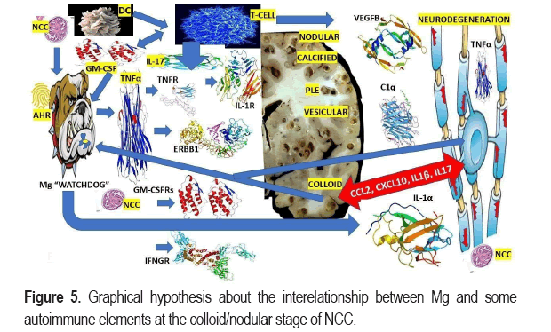
Over the past ten years, most of the investigators have been discussing Mg terminology, mainly "M1 versus M2" or "resting Mg (homeostatic) versus activated Mg", permanently separating Mg into two groups, "good" Mg or "deficient" Mg. However, today it is clear-cut that Mg has more states and functions in CNS disorders, development, ageing, and neuroplasticity than we considered before. To get a better consensus about this issue, Paolicelli et al. organized a group of top-range experts on Mg to analyses the current understanding of Mg states as a dynamic concept and the relevance of addressing Mg function [37]. Based on the previous statement, we have hypothesized that Mg surrounding the cysticercus are permanently moving and scanning the CNS region where they are located with a particular focus on neuronal activity, and soon after, the NCC goes to the colloid state. It reacts according to the characteristic of the neuroimmune system for each location. We also consider that Mg status and function of the activated/M1 are closed related to the sex of the carrier, number and locations of lesions, associated comorbidities, and the capacity of participants in vascular-/glia-/neurogenesis, synaptic modulation, myelination, and delivering soluble factors among other factors at any given CNS topographic region and time as shown in Figure 5.
We have hypothesized that released NCC antigen material during the colloid/nodular stage led to activate Mg cell inducing T-cell activation by myelin autoantigen through IL-17/dendritic cells.
Activated T-cell secret proinflammatory cytokine (IL-17/GM-CSF) inducing Mg to promote neurodegeneration and activate transcription factor NF-kB and expression of transcription factor Aryl Hydrocarbon Receptor (AHR) is downregulated and Mg secret/upregulate TNF-α, C1q, IL-1β, IL-1α, and VEGFB, The Aryl Hydrocarbon Receptor (also known as ahR, AhR, ahr, AH receptor, or dioxin receptor) is a protein transcription factor that regulates gene expression. Tumor Necrotic Factor alpha (TNFα), Interleukin-1 Receptor (IL-1R) (also known as CD121a), This protein is an important mediator involved in many cytokines induced immune and inflammatory responses, the Tumor Necrosis Factor Receptor (TNFRs) is a protein of cytokine receptors characterized by the ability to bind TNFs through extracellular cysteine-rich domain, FMS-Like Tyrosine kinase (FLT1) is a member of Vascular Endothelial Growth Factor Receptor 1 (VEGFR1) protein, encode a receptor tyrosine kinase which is activated by placental-growth factor and VEGF-A/B. The Epidermal growth factor Receptor (ERBB1) (ErbB-1; HER1 in humans) is a transmembrane protein-receptor for extracellular protein ligands and members of the epidermal growth factor family, Granulocyte-Macrophage Colony-Stimulating Factor (GM-CSFR), also known as Colony-Stimulating Factor 2 (CSF2), is a monomeric glycoprotein secreted by endothelial cells, T cells, Mp, mast cells, NK cells, and fibroblasts that functions as a cytokine, Interleukin-17 Receptor (IL17R) is a cytokine receptor binding proinflammatory cytokine interleukin 17A, a member of IL-17 family ligands produced by T helper 17 cells (Th17). The Interferon-Gamma Receptor (IFNGR) protein complex, Interleukin-1 alpha (IL-1α) also known as hematopoietin 1 is a cytokine of the interleukin 1 family responsible for the production of inflammation produced by activated macrophages, as well as neutrophils, epithelial cells, and endothelial cells. The complement component 1q(C1q) is a protein complex involved in the complement system, which is part of the innate immune system, Vascular Endothelial Growth Factor B (VEGFB) is a protein that encoded by the VEGF-B gene belongs to the vascular endothelial growth factor family. The chemokine ligand (CCL2) 2 is also referred to as monocyte chemoattractant protein 1 and small inducible cytokine A2. It is a small cytokine that belongs to the CC chemokine family, which tightly regulates cellular mechanics, C-X-C motif Chemokine Ligand (CXCL10) 10 also known as interferon gamma-induced protein 10 is a small cytokine belong to the CXC chemokine family, located on chromosome 4 and secreted by EC, fibroblast, and monocytes in response to IFN-γ. Its main function are chemoattraction (T-cells, NK cells, DC, monocytes/Mp, antitumor activity, angiogenesis and T-cell adhesion to EC Interleukin-1 beta (IL-1β) also known as leukocytic endogenous mediator, lymphocyte activating factor, leukocytic pyrogen, mononuclear cell factor, among other names is cytokine protein encoded by the IL1B gene and it’s precursor is cleaved by cytosolic caspase 1 to form mature IL-1β, Interleukin 17(IL-17) family is a family of pro-inflammatory cystine knot cytokines, produced by a group of T helper cell known as T helper 17 cell in response to their stimulation with IL-23, Dendritic Cells (DC) are antigen-presenting cells (also named as accessory cell) of the immune system which main function is to process antigen material and present it on the cell surface to the T cells of the immune system. They work as messengers between the innate and adaptive immune systems, Granulocyte-Macrophage Colony-Stimulating Factor (GM-CSF) also called as colony-stimulating factor 2, is a monomeric glycoprotein secreted by EC, T cells, Mp, NK cells, mast cells, and fibroblasts and work as a cytokine, Perilesional edema (PLD).
Some authors described several stages and models of Mg, such as Disease-Associated Mg (DAMs) mainly in connection with Alzheimer's Disease (AD), Mg Neurodegenerative phenotype (MgNd), Interferon-Responsive Mg (IRMg), Activated Responsible Mg (ARMg), Human AD Mg (HAMg), Parkinson's Disease (PD) Mg signature (PDMgS), Mg Inflamed in Multiple Sclerosis (MS) (MgIMS), Amyotrophic Lateral Sclerosis (ALS)-Associated Signature (ALSAS), Glioma-Associated Mg (GAMg), Lipid-Droplet-Accumulating Mg (LDAMg), White Matter-Associated Mg (WMAMg), and Axon Tractassociated Mg (ATAMg). Based on these communications and knowing the pathogenesis of intraparenchymal, intraventricular, subarachnoid, spinal, and optic nerve NCC, we have hypothesized that there are other states/models of Mg in these conditions that might be named Mg NCC instead of M1/M2/resting/active Mg, which also require resident CD4+T cell for adequate maturation/transition and respond to the influence of the B/T lymphocytes, NK cells, and other immune cells changing their molecular profile, morphology, and ultrastructure, motility and function attending to the modifying factors as aforementioned. Future I investigations using the P2RY12 Mg marker might prove the differences between Mg and Mp in NCC and their role in NI/neuroplasticity. Further well-designed investigations could accurately clarify the exact role of Mg in the process of inflammation of neural tissue during the colloid/nodular/fibrotic states of NCC, with an associated entry of leukocytes into the CNS, with or without associated ischemic stroke, with or without ES/Ep, with or without other CNS injury/damage, and under different levels of innate and adaptive immune responses. Probable at that stage, the terminology of neuroinflammation/Mg activation will be replaced by another one.
Recently, Dermitzakis et al. confirmed the cited postulates and highlighted the role of Mg as a tissue resident Mp primary innate immune cells in the brain and its biological role in maintaining homeostasis through removing bacteria and viruses. In our opinion, other foreign particles, including cysticercus debris during the colloid/nodular stage, adopting a malleable morphology characterized by many branches, retracted processes and enlarged ameboid shape (pro-inflammatory phenotype/high phagocytic) and constantly surveillant the CNS parenchyma, but also synapses, cellular death and mediation of neurogenesis following neuronal damage during colloid-NCC, and protection of neural tissue, also developing the adult mammalian brain, releasing trophic factor, pro/anti-inflammatory elements, phagocytosis, synaptic patterning, cell positioning/survival, axonal dynamics, and myelinogenesis [67]. It has been established that Mg is the only tissue-resident Mp that differs from their hematogenous source due to their environment, which is immune-privileged owing to the construction of the BBB [38] supported by the glymphatic system and AQP4/CA/As (IL33) [68]. We also considered that Mg/NCC need TGF-β to keep their capacity of surveillant on surrounding nerve cells and an impact on its development from E14.5., as has been reported by other authors in other pathological conditions [69].
Recently, we commented on the role of micro Ribonucleic Acid (miRNA) in viral infections [70]; now we have hypothesized that the antigens released by cysticercus during the colloid stage of NCC may dysregulated functional miRNA produced by neurons/GC in the brain targeting several proteins, which deregulates neurons/GC signalling, cell death, gene expression, innate neuroimmune response and modulates the messenger RNA (mRNA). We also speculate that miRNA will be a crucial diagnostic element and the therapeutic molecule to curative therapy for many diseases [70]; in cases of NCC, the depletion miR-124 triggers Mg activation. However, we do not have enough evidence to elaborate a confident hypothesis about it and the same biological feature of all Mg states and their function at each cerebral region for each specific management soon.
We assumed that Mg at the perilesional region of NCC-colloid/nodular stage plays a similar role to other innate immune cells do in the skin (Langerhans cells), liver (Kupffer cells), lungs (alveolar macrophages), bone tissue (osteoclasts), cornea/retina (neural-associated macrophages), PNS (endoneurial and epineurial Mp), subdural leptomeningeal Macrophages (MMs), Choroid Plexus Macrophages (CPM), and Perivascular Macrophages (PVM), which are different from perivascular Mg located between two basal laminae (endothelial cells/glia limitans). In summary, we speculated that during the local transforming process of NCC from the vesicular stage to the colloid/nodular stage CNS Associated Mp (CAMs) and other involved immune cells (dendritic cells, T/B lymphocytes, NK cells, monocytes, NKT cells) stay at the CNS border (meninges/choroid plexus). Notwithstanding, Mg has the privilege the stay nearby of the neuron's community as the only immune cells addressing the surveillance of all neuronal activities [71].
Brief comments on apoptosis, pyroptosis, necroptosis, and PANoptosis in NCC
It’s classically accepted that Np is a type of programmed form of inflammatory cell death/ necrosis well differentiated from Apoptosis (Ap) due to a lack of participation in caspase activation from released mitochondria cytochrome C and leakage of cell debris into the ECS. As in NC, at the perilesional region in the colloid/nodular stage, the cells die due to injuries/damage of the neurons and supportive cells instead of the programmed type of death.
Because of limitations for the extension of this manuscript, other aspects related to MG/NCC/IS /ES /Ep/ES were omitted from this article. However, it will be reviewed in subsequent publications. As far as we know, this review is the first done on Mg's role in NCC [38-45]. Further, well-designed investigations will confirm or deny all hypotheses. In addition, they will bring clarification to other aspects that remain obscure such as the final anatomical destination of Mg after being derived from the yolk sac when migrating/infiltrating the developing brain [46-55], to define the proper function of CAMs during development, health and disease to establish a better understanding of the role of Mg/CAM in the pathogenesis of neurodegenerative/ psychiatric/ inflammatory conditions [56-65], and how Mg contribute to the mechanism of nerve repair/scar formation/calcification/ neuroprotection/immune respond during the vesicular/colloid/ nodular/ calcify stages of NCC and identify the therapeutic target elements to provide secure prevention of ES/Ep/SE/IS in NCC with particular emphasis on the management of miRNA/dysbiosis/GC/pericytes safety/oxidative stress/neuron cells protection and prophylaxis of apoptosis among other aims [72-74]. It has been established that the immune system uses several mechanisms to control different pathologies caused by infections and regulated inflammatory responses [33,34,51,66], as we documented before [75].
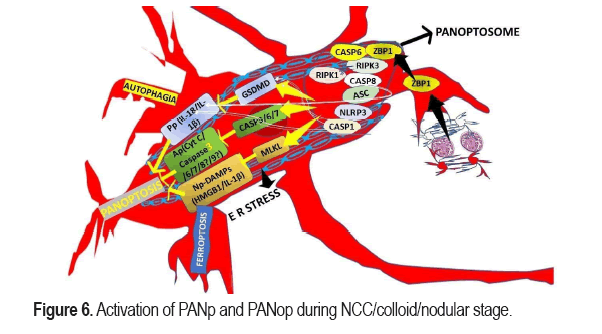
In this statement, we included some zoonotic parasitic brain infections remarkably represented by NCC [5]. Based on other author's publications [76], we considered that the CNS' Mg cell could identify the signals produced by NCC/colloid/nodular antigens over the surrounding cells and respond quickly, producing pro-inflammatory elements and activate Programmed Cell Death (PCD) mechanisms including Apoptosis (Ap), Pyroptosis (Pp), Necroptosis (Np), Autophagy (Aph) and Ferroptosis (Fp). We have hypothesized that according to the number of cysticerci (larva stage) of pig tapeworm in the brain parenchymal, the quality of the host's immune response (pro-inflammatory cytokines/chemokines), the predominant locations of the cysticercus and the prevalent pathological stages of the cyst, then different types of programmed cell death can be seen in isolated or combined presentation of PANoptosis (PANp) which included apoptosis, pyroptosis, and necroptosis to attenuated the severity/consequences of the infection as shown in Figure 6.
We have hypothesized that NCC infection is also sensed by TLRs and innate receptors to elaborated/release NFκB-dependent inflammatory cytokines, (including TNF superfamily) as we commented in previous articles [68], which promote inflammatory signalling through death receptors such as TNFR1, Fas, TRAIL-R, and DR3. Combined death receptors signal and TLRs via adaptor proteins like TRADD, FADD, RIPK1 engage downstream signalling pathways (TAK1 and NF-κB). If TAK1 activity fail, signalling through these receptors leads PCD via RIPK1. We also considered that TAK1-mediated suppression of PANoptosis can be affected by dysfunctional ES mechanism promoting a PANoptosome formation plus a remarkable downstream expression of Ap (CASP3/6/7), Pp (GSDMD), and Np (MLKL) executioners as is illustrated. In this hypothesis we included the adaptor ASC recruiting and activating caspase 1/8 when inflammasome sensors identify their specific parasitic PAMPs. In summary, the process of PANp and the formation of PANop happen after stimulated, ZBP1, RIP1, RIP3, caspase-1, ASC, NLRP3, FADDand caspase-8 form PANop, mediating PANp. We also hypothesized that Caspase-6 is a factor regulating the interaction between RIP3 and ZBP1 in NCC. To deliver a comprehensive hypothesis about the TLR4 signalling pathways in Mg during NCC we could not find supporting evidence on the molecular mechanism for caspase-8 activation. However, we commented about these issues (TLR4) in previous publications [16-20, 68]. Apart from the before-cited PCD, in this hypothesis we included Oxytocic/Ferroptosis (Fp) is another (genetically and biochemically) different type of PCD depending on iron’s CNS and characterized by increasing deposit of lipid peroxides but this issue will be commented in detail in forthcoming articles. In this Figure 6 we summarized the participation of ZBP1 (innate sensor of Influenza A Virus (IAV)) that triggers NI and PANoptosis and ZBP1 activation interacting with caspase-3/6/7, RIPK3, RIPK1, and caspase-8 to assembling the PANoptosome. We have hypothesized that ZBP1-dependent PANoptosome in cases of NCC also engages NLRP3 inflammasome activation and GSDMD-dependent pyroptosis as is illustrated in this figure including activation of caspase-8 and we propose that in NCC the caspase leads to caspase-3-6-7 activation, Ap and cleave GSDMD, while inhibition of caspase-8 cause phosphorylation of MLKL and Np. Because, in NCC there are simultaneously different pathological process according with the number on cysticercus, location, stage of the cysticercus, and local immune response we proposed that PANp is the predominant PCD for massive NCC with fatal outcome.
We assumed that the larva stage of NCC between the vesicular and calcified stages is under severe attack by activated Mg/Mp, activated Ast plus many autoimmune pro-inflammatory elements and the surrounding tissues in high alert before selecting which Ap way to take. We also consider that endogenous Ap (mitochondrial Ap) is involved in this process soon after the protein secretion/excretion mechanism is not good enough to keep the immune response under control. The damage of the outer mitochondrial layer allows the release of cytochrome C (cyto-C), which activate caspase-9, followed by caspase-3, 6, and 7 expressions leading to Ap/PCD. We think that in similar scenarios in which exogenous Ap might predominate because of the interaction of ligands and receptors, sending Ap signals via TNF Receptor 1 Associated via Death Domain (TRADD) and Fas associating protein/novel Death Domain (FADD), then the receptor binds to caspase-8 precursor and its expression activate caspase-8 and later caspase-3, 6, 7 triggering Ap. On the other hand, the RE stress as another way of Ap also contributes to eliciting PANp.
We also speculate that in NCC, both Np and Pp are lytic forms of PCD manipulated by the activation of Mixed Lineage Kinase domain-Like (MLKL) and the pore-forming proteins Gasdermin D (GSDMD), which mediates Pp, release intracellular cytokine and Damage-Associated Molecular Patterns (DAMPs). At the same time, Np happens after Receptor-Interacting serine/threonine-Protein Kinase (RIPK) 3-dependent phosphorylation (MLKL), producing disruption of membrane integrity and MLKL translocation towards plasma membrane as has been proposed for other conditions. PANptosis is the only PCD regulated by the PANoptosome [77] as shown in Figure 6.
In 2019, the terminology of PANoptosis was proposed by Malireddi et al. These authors describe a novel PCD mechanism by the interaction of Ap, Pp, and Np, whose results could not be explained by any of them separately [77,78].
Based on the previous author's experiences, we postulated that regulated cell death (Ap, Pp, and Np) are superimposed among each other and are now grouped under the name of NANp. Figure 5 represents the ER stress as a novel apoptosis regulatory pathway. We suspect t in cases where ER stress is present due to NCC. The system will react with an unfolding protein response, which is activated by: Protein kinase R-like Endoplasmic Reticulum Kinase (PERK), Activating Transcription Factor 6 (ATF6) and Inositol-Requiring Enzyme 1 (IRE1), which are ER stress-integrated proteins [78]. Under different conditions, the same author established that the interaction between adaptor protein TNF Receptor-Associated Factor 2 (TRAF2) and IRE1 triggers the activation of Jun N terminal Kinase (JNK) and promotes activation of pro-apoptosis protein Bcl-2 associated death promoter (Bad) which induce programmed cell death; therefore, we assumed same happens in cases with NCC. Apoptosis can be promoted by C/EBP-Homologous Protein (CHOP) expression upregulating Bcl-2 antagonist/killer (Bak) and pro-apoptosis proteins Bcl-2 associated X protein (Bax). On the other hand, it is well known that PERK activates the translation of ATF4, which can promote C/EBP-Homologous Protein (ER stress protein (CHOP)) expression and promote the phosphorylation of eukaryotic translation Initiation Factor 2α (eIF2α) [79].
Taking into consideration the before cited article, we have hypothesized that RIP3 and Mixed Lineage Kinase domain-Like (MLKL), Receptor-Interacting serine/threonine-Protein kinase 1 (RIP1), and RIP3 regulate necroptosis, and the combination of RIP1/RPI3 produce a complex known as necrosome which is responsible for autophosphorylation. As is illustrated in Figure 5, soon after, RIP3 phosphorylates MLKL protein which activates NLRP3 inflammasome and leads to translocation to the cell membrane and production of MLKL oligomers, causing cell death in patients presenting NCC either. In the past, we tried to explain the radiological differences seen at each NCC stage and correlated it with each feature of cell death programmed independently. Each cell death modality is very strongly linked, and one is regulated by another and vice versa, as reported [80]. Because therapy of Ap inhibitor might suppress Pp and activate Np in animal studies, it has been considered that Pp inhibitor then can suppress Pp and activate Np [81].
Supported by a recent publication made by Shi and colleagues [78], we included in our hypotheses about the relationship between the molecular switch of cell death, also known as caspase-8 (Cp-8) and Ap, Pp (Cp-3, 8), Np as the regulator. At the same time, we keep out of this hypothesis to inflammatory caspases (Cp-1, 4, 5, and 11) because of a need for more confident evidence. However, we keep another relevant molecular regulator of Ap, Pp, and Np, like GSDMD, in our proposal based on its capacity to interact with Ap and Pp [82], and we also included RIP3 protein because the role of regulating cell Ap or cell Np, knowing that its high expression will lead Np pathway. In contrast, the low expression will guide cells to the Ap pathway. Activate RIP3 also release IL-1β via the NLRP3-caspase-1 axis, which serves to support that RIP3 plays an essential role in regulating the balance of Ap, Pp, and Np, as shown in Figure 6.
Based on some results from investigations done with Pseudomonas aeruginosa, it has been reported that cell-death molecules like RIP1 and MLLK were activated in the absence of NLRC4, whereas Cp-1, 3, 7,8 expressions were reduced as another proof of the crucial relationship among Ap, Pp, and Np as we speculate happens in NCC and shown in Figure 6.
In the next paragraph, we will briefly comment on PANopsome (PANop) and Inflammasome (Inf), respectively, and their role in the pathophysiology of NCC, but before that, we will get highly Z-DNA Binding Protein which is a nucleic acid innate immune sensor required for the activation of NLRP3 and PANp which primary function is regulates NCC-patients' immune response. Its principal function is to trigger Ap, Np, and Pp via RIP3, NLR thermal protein domain associated Protein 3 (NLRP3) and Cp-8 expression being a vital component of PANop which is composed of adapters (apoptosis associated speck-like protein containing a CARD (ASC) and FADD), receptors (INfs, ZBP1 and its Zα2 domain), and catalytic effectors (caspase-1, RIP3, RIP1, and caspase-8) [78]. On the other hand, it has well known that ZBP1 has been upregulated in patients infected by SARS-CoV-2 during β-coronavirus infection (leading to cytokine storm) first time reported in South Africa very close to this area [16-18,20]. We also speculate the role of RIP1 regulating PANop for cell death and inflammatory response to released antigens from the colloid/nodular stage of NCC, including some scenarios where RIP1 can be destroyed between colloid and nodular stage and before calcification of the lesion, in which case Yersinia-induce INsm activation/Pp/Ap is abolished while Np is enhanced. It is crucial to highly that yersinia modulate the components of the PANop complex, such as FADD, NLRP3, RIP1, ASC, caspase 8, and RIP3, to control the PANop pathways, as has been reported under different circumstances [20,83]. Another regulatory element of PANop is the Transforming Growth Factor-β (TGF-β)-Activated Kinase 1 (TAK1). From time to time, one of the problems that we afford treating patients with NCC is the collateral cell damage caused during the dying process of the cysticercus responding to the anti-parasitic medication (praziquantel/albendazole) such as ES/SE/NI among other; we are hypothesized that applying therapeutic procedure able to inactivate caspase8/RIP3 or even FADD/RIP3 might protect NC/GC host defence mechanism from cell death and collateral damage. The presence of the before cited INso (a vital component of PANopsome) for Pp and Ap is mandatory. Based on delivered reports under different scenarios, we also considered that almost all the recognized Inflammasome sensors (NLRP3, NLRP1, Pyrin, Absent In Melanoma 2 (AIM2), and NLRC4) are involved in PANop [78] as we represented graphically in Figure 6. From the sensors before-cited, NLRP3 is remarkably important because, after its activation, inflammatory cells die by PANop, or cell death diminishes when its neutralized. We also believe that the most likely elements for increased cell death in NCC are RIP3 and caspase8. Therefore, we considered that in the colloid/nodular stage of NCC, these PANop complexes induce the activation of downstream cell death effector through Ap (caspase-3/7), Pp (caspase-1 and GSDMD), and Np (RIPK3 and MLKL) suggesting it plays a vital role in infections control depending on several cystic lesions, their location, and the quality of host's immune response. In our h hypotheses, we considered that NCC antigens activate Mg/Mp leading to upregulate expression of caspase-3, GSDMD, caspase-1, RIP3, MLKL, IL1β, IL-18, p-MIKLand NLRP3 in Mg/Mp inducing different levels of programmed cells death according to the stage of the cysticercus. Fortunately, all cystic lesions are not at the same stage because all parasites do not invade the CNS at the same time either. Suppose n cases with NCC, the inhibition of TLR9 might cause suppressing MAPK pathway/PANop and better prognosis as has been reported in other pathological processes [81]. In Figure 6, we summarized the process of PANp. Considering that NCC is a gradual pathological process from the beginning with as escalating infection, considering that all cysticerci together do not invade the whole CNS at the same time from the blood flow, considering that all cysticercus are not at the same stage of life or dying process due to natural causes or by pharmacological effect of the anti-parasitic drugs, considering that they are not located at the same place which depend of the laminal/turbulent blood flow and considering that the immune response is not uniform through the CNS based on multiple variations from the brain cortex/ spinal cord to the posterior root ganglion which means all NC/GC do not have the same level of immunological protection all times then we speculate that all NC/GC may have different ways of die responding to different quality/quantity stimulations and activating different signalling pathways in the NC/GC leading to different cell death pathways which justify the terminology of PANop as the preferably way of death in NCC.
One relevant aspect of T solium NCC is the capacity to suppress/evade the host innate immunity function of Mg by releasing an abundant quantity of Excretory/Secretory Proteins (ESPs), which are a heterogeneous group of glycoproteins, proteins and another group of necessary molecules that allow vesicular stage of NCC to survival and remain healthy in the host's CNS under closed surveillance of Mg.
For many years until this relationship (released proteins/host immunity) is broken-down and Mg activation change altogether, this scenario, as we suspected in Binswanger disease and NCC a long time ago [2].
Based on these previous arguments, we hypothesized that at the beginning of the colloid stage and responding urgently to released stimulant antigens, NC/GC preferentially select the swift mode of death identified as Pp according to those cells' death Gene Expression (GDE). In our opinion, the final mode of death selection will depend on the NC/GC's GDE, for examples if GDE-Ap predominates, then the cell will die through Ap, but if the GDE-Pp predominate, those NC/GC may die by Pp and so on. However, we speculate that all PCDM can be present in multifocal colloid/nodular NCC, where many participating factors emerge from the interrelationship between Ap, Pp, and Np, including the inter-regulation among them. Therefore, the predominant PCDM for NCC is PCD accompanied by increased production of microbicidal molecules (reactive oxygen species and nitric oxide), as we commented in previous articles [5,12,12-18,20,39] and gene expression.
According to our proposal, the NCC colloid/nodular stage is tightly associated with PANop as a predominant Regulatory Programmed Cell Death (RPCD). However, another RPCD characterized by iron-dependent lipid peroxidation called Ferroptosis (Fp) might be involved in the pathophysiology of NCC. Fp is associated with tumour progression, tuberculosis, Malaria, cryptococcal meningitis and COVID-19 [84]. Recently we reported a case presenting a comorbidity of NCC/COVID-19, a high level of Ferritin (an essential protein for storing iron) and avoids cell damage caused by Fenton reaction via combining with excess ferrous iron, but that patient presenting a brainstem dysfunction [16]. Due to the tremendous importance of this mechanism in NCC, we will comment on this issue deeply in our following publication, including the underlying mechanism of Fp in cases infected by NCC, the relationship between Fp and autophagia, the role of selenium, cysteine, GPX4, Glutathione metabolism plus other Fp inhibitors, iron metabolism, lipid metabolism, energy stress, Damage Associated Molecular Patterns (DAMPs), and Fp/Mp, Fp/neutrophil trying to bridge Fp and NCC among other aspects.
Brief comments on somatic NC reprogramming in NCC
Recently, some authors have proposed that Somatic Cell Reprogramming (SCR) is an alternative way to obtain NC based on novel techniques used to reprogram differentiated and mature cells into NC [85].
Because these neural stem cells are remarkably scanty, they cannot restore the large numbers of neurons lost during massive NCC. However, they might do it in cases with limited CNS cysticercosis lesions. However, the exact number of cysts has yet to be identified. A similar situation has been confirmed in adult neural stem cells after cortical IS stroke lesions lead to reactive Ast [86] these two experiments showed that the nuclei of adult cells could be reprogrammed so that they exhibited cellular characteristics like those of fertilized eggs. In a later study, Japanese scientist Tommo Nakayama transplanted the nuclei of adult rat skin cells into embryonic stem cells. This study showed that the nuclei of adult cells can be reprogrammed to produce further embryonic stem cells. Moreover, these stem cells could differentiate into different types of cells [12]. In 2006, scientist Shinya Yamanaka successfully transformed adult mouse skin cells into pluripotent stem cells capable of differentiating into multiple cell types by subjecting them to genetic transformations and cultures [13], which led to his winning the Nobel Prize. This achievement was considered a revolutionary advancement in cell biology and medicine and laid the foundation for subsequent research. In subsequent studies, scientists discovered that it was possible to obtain mature neurons by converting somatic cells into stem cells and then differentiating them into neural precursor cells with a dozen transcription factors [14-17].
Indeed, ectopic overexpression of specific neuronal determinants can reprogram non-neuronal cells directly into fully functional neurons in vivo and in vitro [18-20]. Since this pioneering work, researchers have explored how to exploit transcription factors, microRNAs (miRNAs), and gene silencing to reprogram highly differentiated, mature cells into neurons for therapeutic purposes. Generally, the rationale is to suppress the expression of genes subserving the current cell phenotype while activating genes that give rise to the target cell phenotype [21,22].
Sophisticated techniques and tools are now available to reprogram somatic cells into neurons. Multiple types of somatic cells have been investigated in neural reprogramming. Reprogramming is easier and more effective when the somatic cell phenotype resembles the target neuron lineage. Astrocytes are currently thought to be ideal candidates for neural repair. Human fi oocytes are also other ideal sources of reprogramming in this field because they are relatively easy to obtain. Many types of cells from other tissues and embryonic cortices have been involved in the mechanism of Reprogramming of Somatic Cells (RSC), mainly ectodermal cell types such as fibroblast, Osteoclast Like Cell (OLC), Ast, keratinocytes, NC and Pc [87]. These authors established that this is happening because progenitor cells and embryonic cells can differentiate into cells with similar genealogical sources; therefore, neuronal ectoderm can produce Pc, Ast and fibroblasts, but Ast are the best candidates for neuro regenerative reprogramming on top of Mg [87] that is why we hypothesized that from nodular to the calcified stage most of the new neurons replacing the NC lost during the PCD mechanism are from Ast/RSC.
Our last hypothesis will try to answer the last research question regarding another mechanism for NC protection not mentioned before. For the past twenty-six years, we have seen many times several patients with complete recovery from multiple and bilateral vesicular/colloid/nodularfibrotic-NCC without headache, ES/Ep or other clinical sequels who remain entirely asymptomatic, presenting just CNS calcifications or even calcified NCC. Apart from the somatic NC reprogramming, which guarantees new NC replacement from ectodermic cells, and NG reprogramming from neuronal ectoderm, more evidence is necessary to explain how so many damaged/Ap/Np/PANp NC are going to be replaced. Then, we hypothesized that other process works from the beginning of the NCC infection before the PCD/RSC programmed supporting NC protection, limiting the number of NC/NG cellsfrom the pericystic area and how the immune response is improved gradually leading to a better outcome and reduced mortality rate. We have hypothesized that Mg (first line of defence) can potentiate its response by a nonspecific attack against new cysts invading CNS tissues following new ingestion of eggs/proglottids of NCC from contaminated water/food through a way of the innate type of immunological memory also known as trained immunity like a fast-acting, nonspecific memory, also considered as a new weapon in the fight against infectious diseases, as has been nominated by other authors [88]. We expect that chemical/physical obstacles besides myeloid/innate lymphoid cells mediate these responses to antigens released from the vesicular/colloid stage. We also speculate that Mg has an innate immunological memory based on the innate immune cells and immune-GC, which are expressed soon after they expose to NCC antigens, as has been postulated by other authors for other situations [89]. We also speculate that soon after the first exposure of CNS to NCC/colloid antigens, two types of altered innate immune response can happen depending on the quantity and location of the cystic lesions, such as anti-inflammatory response state, also named as tolerance or pro-inflammatory response, also called trained immunity because its trains challenger cells and triggers an immune response. Other estimators have established that innate immune cells change the epigenetic landscape chromatin structure during this process, decreasing DNA methylation, activating pro-inflammatory genes, and increasing histone methylation and acetylation marks [90].
Conclusion
The NCC can suppress Th1 and Th2 responses by ES products preventing tissue damage during the vesicular stage and allowing the larva stage of NCC to survive for many years in humans. We have hypothesized that the relationship between the chronic stimulation causing by the different arrival of cysticercus to the CNS and the trained immunity in endemic regions for NCC provides an ideal scenario to attenuate the disease while novel therapies to kill the parasite with the absolute absence of side effect and complications. As far as we know, no previous review of this issue has been published, and other investigations are necessary to confirm/deny/modify the previously delivered hypotheses.
Declaration
Consent for publication
We obtained the written informed consent for publication from our patient, including laboratory results. All information is fully available for any interested reader by request.
Ethical approval
The WSU/NMAH Ethical Committee did not request ethical approval for this study.
Competing interest
The authors declare that they performed this study without any commercial, financial, or otherwise relationships able to construe a potential conflict of interest.
Funding
All authors declare that they did not receive financial aid or collaboration that could have influenced the results reported in this paper.
Author’s contributions
Study concept and design: HFS. Data collection from searched literature: HFS. Analysis of the obtained data was done by HFS plus the first and final draft of this paper. Manuscript was revised by HFS. Manuscript writing process: HFS. The author has approved this version for publication.
Declaration of anonymity
The authors certified that they did not mention names, initials, and other identity issues of any patient. Therefore, a complete anonymity is guaranteed.
Availability of data and materials
All data supporting this study are available on reasonable request from the corresponding author.
Acknowledgment
Special thanks to Dr Lourdes de Fatima Ibañz Valdés and Dr Sibi Joseph for their participation in the management of our patients and Professor Thozama Dubula, The Dean of Faculty of Health Sciences of Walter Sisulu University and Dr Kulile Moetkhesi for their unconditional support.
References
- Foyaca-Sibat, Humberto, and LdeF Ibanez Valdes. "Pseudoseizures and Epilepsy in Neurocysticercosis." Rev Electron Biomed 2 (2003):79-87.
- Foyaca-Sibat, Humberto, and LdeF Ibanez Valdes. "Vascular Dementia Type Binswanger's Disease in Patients with Active Neurocysticercosis." Rev Electron Biomed 1 (2003):32-42.
- Foyaca-Sibat, Humberto, and LdeF Ibanez Valdes. "Insular Neurocysticercosis:Our Finding and Review of the Medical Literature." Intern J Neurol 5 (2006):31-35.
- Foyaca-Sibat, Humberto, Linda D Cowan, Helene Carabin, and Irene Targonska, et al. "Accuracy of Serological Testing for the Diagnosis of Prevalent Neurocysticercosis in Outpatients with Epilepsy, Eastern Cape Province, South Africa." PLoS Negl Trop Dis 3 (2009):e562.
- Foyaca-Sibat, Humberto, LdeF Ibanez Valdes, and J Moré-Rodríguez. "Parasitic Zoonoses of the Brain:Another Challenger." Intern J Neurol 12 (2010):9-14.
- Foyaca-Sibat, Humberto, and LdeF Ibanez Valdes. "Treatment of Epilepsy Secondary to Neurocysticercosis". Intech Open (2011).
- Foyaca-Sibat, Humberto, and LdeF Ibanez Valdes. "Clinical Features of Epilepsy Secondary to Neurocysticercosis at the Insular Lobe." Intech Open (2011).
- Foyaca-Sibat, Humberto. "Epilepsy Secondary to Parasitic Zoonoses of the Brain." Intech Open (2011).
- Foyaca-Sibat, Humberto, M Salazar-Campos, and LdeF Ibanez Valdes. "Cysticercosis of the Extraocular Muscles. Our Experience and Review of the Medical Literature." Intern J Neurol 14 (2012).
- Foyaca-Sibat, Humberto, and LdeF Ibanez Valdes. "Introduction to Cysticercosis and Its Historical Background." Intech Open (2013).
- Foyaca-Sibat, Humberto, and LdeF Ibanez Valdes. "What is a Low Frequency of the Disseminated Cysticercosis Suggests that Neurocysticercosis is Going to Disappear?." Intech Open (2013).
- Foyaca-Sibat, Humberto, and LdeF Ibanez Valdes. "Uncommon Clinical Manifestations of Cysticercosis." Intech Open 8 (2013):199-233.
- Foyaca-Sibat, Humberto, and LdeF Ibanez Valdes. "Psychogenic Nonepileptic Seizures in Patients Living with Neurocysticercosis." Intech Open (2018).
- Foyaca-Sibat, Humberto, and LdeF Ibañez-Valdés. "Subarachnoid Cysticercosis and Ischaemic Stroke in Epileptic Patients." Intech Open (2018):153.
- Noormahomed, Emilia Virgínia, Noémia Nhancupe, Jerónimo Mufume, and Robert T Schooley, et al. "Neurocysticercosis in Epileptic Children:An Overlooked Condition in Mozambique, Challenges in Diagnosis, Management and Research Priorities." EC Microbiol 17 (2021):49-56.
- Humberto Foyaca Sibat. "Racemose Neurocysticercosis Long COVID and Brainstem Dysfunction:A Case report and Systematic Review”. Clin Schizophr Relat Psychoses 15S (2021).
- Foyaca-Sibat, Humberto. "Neurocysticercosis, Epilepsy, COVID-19 and a Novel Hypothesis:Cases Series and Systematic Review." Clin Schizophr Relat Psychoses 15 (2021):1-13.
- Foyaca-Sibat, Humberto. "People Living with HIV and Neurocysticercosis Presenting Covid-19:A Systematic Review and Crosstalk Proposals." Clin Schizophr Relat Psychoses 15 (2021):1-9.
- Foyaca-Sibat, Humberto, and LdeF Ibanez Valdes. "Novel Hypotheses on the Role of Oligodendrocytes in Neurocysticercosis. Comprehensive Review". Clin Schizophr Relat Psychoses 17 (2023).
- Foyaca-Sibat, Humberto. "Comorbidity of Neurocysticercosis, HIV, Cerebellar Atrophy and SARS-Cov-2:Case Report and Systematic Review." Clin Schizophr Relat Psychoses 15 (2021):1-6.
- Del Rio-Romero, A H, H Foyaca-Sibat, LdeF Ibanez-Valdes, and E Vega-Novoa. "Prevalence of Epilepsy and General Knowledge about Neurocysticercosis at Nkalukeni Village, South Africa." Intern J Neurol 3 (2005):1-6.
- Del Rio-Romero, A H, H Foyaca-Sibat, LdeF Ibanez-Valdes, and E Vega-Novoa. "Neuroepidemiological Survey for Epilepsy and Knowledge about Neurocysticercosis at Ngqwala Location, South Africa." Intern J Neurol 4 (2004):1-6.
- Foyaca-Sibat, Humberto, A Del Rio-Romero, and L. Ibanez-Valdes. "Prevalence of Epilepsy and General Knowledge about Neurocysticercosis at Ngangelizwe Location, South Africa." Intern J Neurol 4 (2005):23-37.
- Del Rio-Romero, A H, and H Foyaca-Sibat. "Epidemiological Survey about Socio-Economic Characteristic of Mpindweni Location, South Africa." Intern J Neurol 4 (2005):18-26.
- Foyaca-Sibat, Humberto, and LdeF Ibanez Valdes. "Refractory Epilepsy in Neurocysticercosis." Intern J Neurol 5 (2005):1-6.
- Foyaca-Sibat, Humberto, and LdeF Ibanez Valdes. "Insular Neurocysticercosis:Our Finding and Review of the Medical Literature." Intern J Neurol 5 (2006):31-35.
- Foyaca-Sibat, Humberto, and LdeF Ibanez Valdes. "Meningeal Lymphatic Vessels and Glymphatic System in Neurocysticercosis. A Systematic Review and Novel Hypotheses." Clin Schizophr Relat Psychoses 17 (2023):1-11.
- Foyaca-Sibat, Humberto, and LdeF Ibanez Valdes. "Co-morbidity of Spinal Cord Neurocysticercosis and Tuberculosis in a HIV-Positive Patient." Intern J Neurol 7 (2007):5-10.
- Foyaca-Sibat, Humberto, and LdeF Ibanez Valdes. "The Role of Pericytes in Neurocysticercosis:Comprehensive Review and Novel Hypotheses." Clin Schizophr Relat Psychoses 17 (2023):1-12.
- Ludwig, Parker E, and Joe M Das. "Histology, Glial Cells." StatPearls (2022).
- Wang, Jie, Wenbin He, and Junlong Zhang. "A Richer and More Diverse Future for Microglia Phenotypes." Heliyon 9 (2023):e14713.
- Foyaca-Sibat, Humberto, and LdeF Ibanez Valdes. "Introduction to Novel Motor Neuron Disease." Intech Open (2019).
- Hanani, Menachem, and Alexei Verkhratsky. "Satellite Glial Cells and Astrocytes, a Comparative Review." Neurochem Res 46 (2021):2525-2537.
- Lee, Ji Hwan, and Woojin Kim. "The Role of Satellite Glial Cells, Astrocytes, and Microglia in Oxaliplatin-Induced Neuropathic Pain." Biomedicines 8 (2020):324.
- de, Shrutokirti, Donn van Deren, Eric Peden, and Matt Hockin, et al. "Two Distinct Ontogenies Confer Heterogeneity to Mouse Brain Microglia." Development 145 (2018):dev152306.
- Kohno, Keita, Ryoji Shirasaka, Kohei Yoshihara, and Satsuki Mikuriya, et al. "A Spinal Microglia Population Involved in Remitting and Relapsing Neuropathic Pain." Science 376 (2022):86-90.
- Paolicelli, Rosa C, Amanda Sierra, Beth Stevens, and Marie-Eve Tremblay, et al. "Microglia States and Nomenclature:A Field at its Crossroads." Neuron 110 (2022):3458-3483.
- Li, Shuailong, Isa Wernersbach, Gregory S. Harms, and Michael KE Schäfer. "Microglia Subtypes Show Substrate-and Time-Dependent Phagocytosis Preferences and Phenotype Plasticity." Front Immunol 13 (2022):945485.
- Wan, Teng, Yunling Huang, Xiaoyu Gao, and Wanpeng Wu, et al. "Microglia Polarization:A Novel Target of Exosome for Stroke Treatment." Front Cell Dev Biol 10 (2022):842320.
- Ransohoff, Richard M. "A Polarizing Question:Do M1 and M2 Microglia Exist?." Nat Neurosci 19 (2016):987-991.
- Guo, Shenrui, Hui Wang, and Yafu Yin. "Microglia Polarization from M1 to M2 in Neurodegenerative Diseases." Front Aging Neurosci 14 (2022):815347.
- Michelucci, Alessandro, Tony Heurtaux, Luc Grandbarbe, and Eleonora Morga, et al. "Characterization of the Microglial Phenotype Under Specific Pro-Inflammatory and Anti-Inflammatory Conditions:Effects of Oligomeric and Fibrillar Amyloid-β." J Neuroimmunol 210 (2009):3-12.
- Mills, Charles D, Kristi Kincaid, Jennifer M Alt, and Michelle J Heilman, et al. "M-1/M-2 Macrophages and the Th1/Th2 Paradigm." J Immunol 164 (2000):6166-6173.
- Butovsky, Oleg, Mark P Jedrychowski, Craig S Moore, and Ron Cialic, et al. "Identification of a Unique TGF-β-Dependent Molecular and Functional Signature in Microglia." Nat Neurosci 17 (2014):131-143.
- Martinez, Fernando O, and Siamon Gordon. "The M1 and M2 Paradigm of Macrophage Activation:Time for Reassessment." F1000Prime Rep 6 (2014).
- Streit, Wolfgang J, Manuel B Graeber, and Georg W Kreutzberg. "Functional Plasticity of Microglia:A Review." Glia 1 (1988):301-307.
- Acarin, Laia, Jose M Vela, Berta González, and Bernardo Castellano. "Demonstration of Poly-N-Acetyl Lactosamine Residues in Ameboid and Ramified Microglial Cells in Rat Brain by Tomato Lectin Binding." J Histochem Cytochem 42 (1994):1033-1041.
- Kitamura, Tadahisa, Toshihiko Miyake, and Setsuya Fujita. "Genesis of Resting Microglia in the Gray Matter of Mouse Hippocampus." J Comp Neurol 226 (1984):421-433.
- Tremblay, Marie-Ève, Cynthia Lecours, Louis Samson, and Víctor Sánchez-Zafra, et al. "From the Cajal Alumni Achúcarro and Río-Hortega to the Rediscovery of Never-Resting Microglia." Front Neuroanat 9 (2015):45.
- Tremblay, Marie-Ève. "The Role of Microglia at Synapses in the Healthy CNS:Novel Insights from Recent Imaging Studies." Neuron Glia Biol 7 (2011):67-76.
- Borst, Katharina, Anaelle Aurelie Dumas, and Marco Prinz. "Microglia:Immune and Non-Immune Functions." Immunity 54 (2021):2194-2208.
- Böttcher, Chotima, Stephan Schlickeiser, Marjolein AM Sneeboer, and Desiree Kunkel, et al. "Human Microglia Regional Heterogeneity and Phenotypes Determined by Multiplexed Single-Cell Mass Cytometry." Nat Neurosci 22 (2019):78-90.
- Kalkman, Hans O, and Dominik Feuerbach. "Microglia M2A Polarization as Potential Link between Food Allergy and Autism Spectrum Disorders." Pharmaceuticals (Basel) 10 (2017):95.
- Yunna, Chen, Hu Mengru, Wang Lei, and Chen Weidong. "Macrophage M1/M2 Polarization." Eur J Pharmacol 877 (2020):173090.
- Jander, Sebastian, and Guido Stoll. "Strain‐Specific Expression of Microglial Keratan Sulfate Proteoglycans in the Normal Rat Central Nervous System:Inverse Correlation with Constitutive Expression of Major Histocompatibility Complex Class II Antigens." Glia 18 (1996):255-260.
- Tränkner, Dimitri, Anne Boulet, Erik Peden, and Richard Focht, et al. "A Microglia Sublineage Protects from Sex-Linked Anxiety Symptoms and Obsessive Compulsion." Cell Rep 29 (2019):791-799.
- Young, Kimberly, and Helena Morrison. "Quantifying Microglia Morphology from Photomicrographs of Immunohistochemistry Prepared Tissue Using ImageJ." J Vis Exp 136 (2018):e57648.
- Arioz, Burak I, Bora Tastan, Emre Tarakcioglu, and Kemal Ugur Tufekci, et al. "Melatonin Attenuates LPS-Induced Acute Depressive-Like Behaviors and Microglial NLRP3 Inflammasome Activation through the SIRT1/Nrf2 Pathway." Front Immunol 10 (2019):1511.
- Xu, Jing, Zhouqing Chen, Fang Yu, and Huan Liu, et al. "IL-4/STAT6 Signaling Facilitates Innate Hematoma Resolution and Neurological Recovery after Hemorrhagic Stroke in Mice." Proc Natl Acad Sci USA 117 (2020):32679-32690.
- Subedi, Lalita, Jae Hyuk Lee, Silvia Yumnam, and Eunhee Ji, et al. "Anti-Inflammatory Effect of Sulforaphane on LPS-Activated Microglia Potentially through JNK/AP-1/NF-Κb Inhibition and Nrf2/HO-1 Activation." Cells 8 (2019):194.
- Stratoulias, Vassilis, and Tapio I. Heino. "MANF Silencing, Immunity Induction or Autophagy Trigger an Unusual Cell Type in Metamorphosing Drosophila Brain." Cell Mol Life Sci 72 (2015):1989-2004.
- Kisucká, Alexandra, Katarína Bimbová, Mária Bačová, and Ján Gálik, et al. "Activation of Neuroprotective Microglia and Astrocytes at the Lesion Site and in the Adjacent Segments is Crucial for Spontaneous Locomotor Recovery after Spinal Cord Injury." Cells 10 (2021):1943.
- Zhou, Dandan, Lei Ji, and Youguo Chen. "TSPO Modulates IL-4-Induced Microglia/Macrophage M2 Polarization via PPAR-γ Pathway." J Mol Neurosci 70 (2020):542-549.
- Hardeland, Rüdiger. "Melatonin and Microglia." Int J Mol Sci 22 (2021):8296.
- Tsai, Cheng-Fang, Guan-Wei Chen, Yen-Chang Chen, and Ching-Kai Shen, et al. "Regulatory Effects of Quercetin on M1/M2 Macrophage Polarization and Oxidative/Antioxidative Balance." Nutrients 14 (2021):67.
- Louboutin, Jean-Pierre, and David S Strayer. "Relationship between the chemokine Receptor CCR5 and Microglia in Neurological Disorders:Consequences of Targeting CCR5 on Neuroinflammation, Neuronal Death and Regeneration in a Model of Epilepsy." CNS Neurol Disord Drug Targets 12 (2013):815-829.
- Dermitzakis, Iasonas, Maria Eleni Manthou, Soultana Meditskou, and Marie-Ève Tremblay, et al. "Origin and Emergence of Microglia in the CNS-An Interesting (Hi) Story of an Eccentric Cell." Curr Issues Mol Biol 45 (2023):2609-2628.
- Foyaca-Sibat, Humberto, and LdeF Ibanez Valdes. "Meningeal Lymphatic Vessels and Glymphatic System in Neurocysticercosis. A Systematic Review and Novel Hypotheses." Clin Schizophr Relat Psychoses 17 (2023):1-11.
- Prinz, Marco, Takahiro Masuda, Michael A Wheeler, and Francisco J Quintana. "Microglia and Central Nervous System-Associated Macrophages-From Origin to Disease Modulation." Annu Rev Immunol 39 (2021):251-277.
- Foyaca-Sibat, Humberto, Sibi Sebastian Joseph, and LdeF Ibanez Valdes. "Multicommodity of Human Immunodeficiency Virus, Human Herpesvirus Seven and Polyneuropathy." Clin Schizophr Relat Psychoses 17 (2023):1-11.
- Prinz, Marco, and Josef Priller. "The Role of Peripheral Immune Cells in the CNS in Steady State and Disease." Nat Neurosci 20 (2017):136-144.
- Hanisch, Uwe-Karsten, and Helmut Kettenmann. "Microglia:Active Sensor and Versatile Effector Cells in the Normal and Pathologic Brain." Nat Neurosci 10 (2007):1387-1394.
- Tremblay, Marie-Eve, Charlotte Madore, Maude Bordeleau, and Li Tian, et al. "Neuropathobiology of COVID-19:The Role for Glia." Front Cell Neurosci 14 (2020):592214.
- Sierra, Amanda, Marie-Ève Tremblay, and Hiroaki Wake. "Never-Resting Microglia:Physiological Roles in the Healthy Brain and Pathological Implications." Front Cell Neurosci (2014):1-2.
- Foyaca-Sibat, Humberto. "Bilateral Putaminal Haemorrhage and Blindness in Times of the Coronavirus Pandemic and Dysbiosis:Case Report and Literature Review." Clin Schizophr Relat Psychoses 15 (2021):1-14.
- Place, David E, SangJoon Lee, and Thirumala-Devi Kanneganti. "PANoptosis in Microbial Infection." Curr Opin Microbiol 59 (2021):42-49.
- Malireddi, RamaKrishna, Prajwal Gurung, Sannula Kesavardhana, and Parimal Samir, et al. "Innate Immune Priming in the Absence of TAK1 Drives RIPK1 Kinase Activity–Independent Pyroptosis, Apoptosis, Necroptosis, and Inflammatory Disease." J Exp Med 217 (2020):e20191644.
- Shi, Chunxia, Pan Cao, Yukun Wang, and Qingqi Zhang, et al. "PANoptosis:A Cell Death Characterized by Pyroptosis, Apoptosis, and Necroptosis." J Inflamm Res 16 (2023):1523-1532.
- Yang, Yuan, Lian Liu, Ishan Naik, and Zachary Braunstein, et al. "Transcription Factor C/EBP Homologous Protein in Health and Diseases." Front Immunol 8 (2017):1612.
- Gullett, Jessica M, Rebecca E Tweedell, and Thirumala-Devi Kanneganti. "It’s all in the PAN:Crosstalk, Plasticity, Redundancies, Switches, and Interconnectedness Encompassed by Panoptosis Underlying the Totality of Cell Death-Associated Biological Effects." Cells 11 (2022):1495.
- Zhou, Ruixi, Junjie Ying, Xia Qiu, and Luting Yu, et al. "A New Cell Death Program Regulated by Toll-Like Receptor 9 through P38 Mitogen-Activated Protein Kinase Signaling Pathway in a Neonatal Rat Model with Sepsis Associated Encephalopathy." Chin Med J (Engl) 135 (2022):1474-1485.
- Taabazuing, Cornelius Y, Marian C Okondo, and Daniel A Bachovchin. "Pyroptosis And Apoptosis Pathways Engage in Bidirectional Crosstalk in Monocytes and Macrophages." Cell Chem Biol 24 (2017):507-514.
- Malireddi, RamaKrishna, Sannula Kesavardhana, Rajendra Karki, and Balabhaskararao Kancharana, et al. "RIPK1 Distinctly Regulates Yersinia-Induced Inflammatory Cell Death, PANoptosis." Immunohorizons 4 (2020):789-796.
- Xiao, Leyao, Huanshao Huang, Shuhao Fan, and Biying Zheng, et al. "Ferroptosis: A Mixed Blessing for Infectious Diseases." Front Pharmacol 13 (2022):992734.
- Chen, Jiafeng, Lijuan Huang, Yue Yang, and Wei Xu, et al. "Somatic Cell Reprogramming for Nervous System Diseases: Techniques, Mechanisms, Potential Applications, and Challenges." Brain Sci 13 (2023):524.
- Faiz, Maryam, Nadia Sachewsky, Sergio Gascón, and KW Annie Bang, et al. "Adult Neural Stem Cells from the Subventricular Zone give Rise to Reactive Astrocytes in the Cortex after Stroke." Cell Stem Cell 17 (2015):624-634.
- Matsuda, Taito, Takashi Irie, Shutaro Katsurabayashi, and Yoshinori Hayashi, et al. "Pioneer Factor Neurod1 Rearranges Transcriptional and Epigenetic Profiles to Execute Microglia-Neuron Conversion." Neuron 101 (2019):472-485.
- Dagenais, Amy, Carlos Villalba-Guerrero, and Martin Olivier. "Trained Immunity: A “New” Weapon in the Fight against Infectious Diseases." Front Immunol 14 (2023):1147476.
- Netea, Mihai G, Jorge Domínguez-Andrés, Luis B Barreiro, and Triantafyllos Chavakis, et al. "Defining Trained Immunity and Its Role in Health and Disease." Nat Rev Immunol 20 (2020):375-388.
- van der Heijden, Charlotte DCC, Marlies P Noz, Leo AB Joosten, and Mihai G Netea, et al. "Epigenetics and Trained Immunity." Antioxid Redox Signal 29 (2018):1023-1040.
Citation: Valdes, Lourdes Fatima de Ibanez and Humberto Foyaca Sibat. “Novel Hypotheses on the Role of Microglia and PANoptosis in Neurocysticercosis.” Clin Schizophr Relat Psychoses 17 (2023).
Copyright: © 2023 Valdes LFI, et al. This is an open-access article distributed under the terms of the Creative Commons Attribution License, which permits unrestricted use, distribution, and reproduction in any medium, provided the original author and source are credited. This is an open access article distributed under the terms of the Creative Commons Attribution License, which permits unrestricted use, distribution, and reproduction in any medium, provided the original work is properly cited.
